Mammogram

What is a Mammogram?

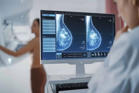
Mammograms are a specific type of imaging that uses a low-dose x-ray system to examine breasts and is used to aid in the early detection and diagnosis of breast diseases in women.

It is considered the gold standard for early detection of breast cancer, capable of identifying abnormalities such as masses, distortions or microcalcifications that may not be detectable through physical examination alone. Mammography is highly effective, often detecting breast cancers long before they can be found by clinical examination, ultrasound, or other methods.

How is the Mammogram procedure performed?
Mammography is performed on an outpatient basis. During mammography, a specially qualified radiographer will position your breast in the mammography unit. Your breast will be placed on a special platform and compressed by a clear plastic plate.

The radiographer will gradually compress your breast.

How is the Mammogram procedure performed?

Mammography is performed on an outpatient basis. During mammography, a specially qualified radiographer will position your breast in the mammography unit. Your breast will be placed on a special platform and compressed by a clear plastic plate.
The radiographer will gradually compress your breast.

Before your Mammogram:

  • It is best to schedule your mammogram for a time when your breasts are least likely to be tender, which is often the week before or after your menstrual period.
  • Please arrive 10 to 15 minutes before your appointment time to complete the paperwork.
  • Bring along any previous mammogram images so that the radiologist can compare them to your new images.
  • You will be asked to complete a questionnaire before your mammogram is done.
  • A cotton gown will be provided for you to wear during the examination.
  • Do not use any roll on, talcum powder or deodorant on the day of your examination.
  • The examination takes approximately 15 minutes.

Are mammograms safe?

  • The benefits of mammography outweigh any possible harm from the radiation exposure. Modern machines use low radiation doses to get breast x-rays that are high in image quality.
  • If there’s any chance you might be pregnant, let your health care provider and x-ray technologist know. Although the risk to the fetus is likely very small, screening mammograms aren’t routinely done in pregnant women.

Why not just an ultrasound?

  • Ultrasound is limited as a screening method as it may not detected small areas of abnormality but is excellent as a examination directed to an abnormality seen on mammogram.
  • However in patients younger than 40 ultrasound is the method of choice to evaluate an abnormal area in the breast.

During your Mammogram

  • Four x-ray images will be taken of your breasts.
  • X-rays will be taken while standing.
  • Your breast will be placed on a plate and compressed by a clear plastic plate in order to spread out the breast tissue.
  • The X-ray unit will move above your head while the images are taken.

After your Mammogram

Sometimes an ultrasound is needed to visualize any abnormalities in the breast tissue. If this is the case, it will be arranged at the time of the Mammogram. If there is any reason for concern, our Radiologist and Mammographers will discuss this with you and any further investigations that are necessary will be explained to you and then booked.

Other Services

Our Women’s Wellness forms part of PRECISION IMAGING. True to its name, we specialize in female health, and offer mammography, ultrasound and bone density scans.

As much of our focus is on women’s health, our Women’s Wellness Centre was established to provide a dedicated area for ladies where they can feel comfortable and enjoy privacy while receiving top quality healthcare.

    • Ultrasound

      Ultrasound

      Ultrasound is a safe, non-invasive, painless & radiation free examination that uses high-frequency sound waves to create images of the body’s internal structures such as organs and blood vessels.

    • X-Ray

      X-Ray

      X-rays are a safe, painless and quick procedure that creates pictures of the inside of your body, usually the bones.

    • Bone Density

      Bone Density

      Bone Density Scanning, also known as Dual-Energy X-Ray Absorptiometry (DEXA) or Bone Densitometry, is a safe low dose X-ray technique used to produce images of the bones.

Precision Imaging

Typically replies within a day